Retinal Vein Occlusion
by Dr Rupal Morjaria
The retina is the light sensitive layer at the back of the eye – like a film in a camera. A healthy retina is important to help process a clear image.
The retina is the light sensitive layer at the back of the eye- like a film in a camera. A healthy retina is important to help process a clear image. Any problems in the retina, particularly the central area called the macula, result in a reduction of vision.
A retinal vein occlusion results in sudden loss of part or all of the vision in the affected eye. It is caused be a blockage in the blood vessels to the retina by a clot (thrombus). The clot can cause a central vein occlusion (whole retina affected), a hemi-vein occlusion (half of retina affected) or a branch vein occlusion (part of retina affected). The effect of a retinal vein occlusion depends on how severe the initial blockage is.
Most people who get a vein occlusion may develop swelling at the back of the eye (called macula oedema) and starvation of of oxygen and nutrients to parts of the retina (called ischaemia). Macular oedema and retinal ischaemia can result in significant visual loss. The presence of macular oedema and ischaemia requires treatment, in the form of laser to the retina and injections to the eye. A very small proportion of people may not need treatment but will need to be carefully monitored to make sure they do not develop further complications.
Ischaemia can occur immediately, a few months or even years after the blockage. When ischaemia does occur, this requires urgent treatment to prevent new retinal blood vessels developing (retinal neovascularisation). These new blood vessels can cause bleeding in the eye resulting in sudden loss of vision or can cause the pressure to build up excessively in the eye resulting in severe pain (rubeotic glaucoma).
People who have suffered from a retinal vein occlusion may therefore require long term monitoring by their Ophthalmologist. Dr Morjaria has expertise in the management of all aspects or retinal vein occlusion and will be able to fully assess the condition, and advise on the treatment and further follow up of the condition to minimise any visual loss or pain.
What will happen to my vision?
Vision following a retinal vein occlusion can vary considerably. Some people experience a complete loss of vision whilst others may get patchy visual loss. Early diagnosis and treatment is key to preventing visual loss. The prognosis depends on your initial vision. Studies have shown that 80% of people who initially present with very poor vision have a lower chance of improvement compared to those who present with a less than 2 lines drop in vision or better. These people can get a complete return of their vision with minimal or no treatment.
What caused my retinal vein occlusion?
The most common cause is a thrombus (blood clot) which blocks the blood vessel supplying the retina. Other conditions that cause inflammation of the blood vessel can also cause this, such as vasculitis.
Risk factors for developing a retinal vein occlusion?
Known high blood pressure or undiagnosed high blood pressure
High cholesterol (>6.5 mmol/l)
Diabetes mellitus
Glaucoma
Blood clotting abnormalities including thrombophilia
Age
Smoking
Rare conditions such as Behçets disease, polyarteritis nodusa, granulomatosis with polyangiitis, acromegaly, Cushing’s syndrome, hypothyroidism
How is the diagnosis made?
Dr Morjaria is a retinal specialist who will examine your eyes to help assess the extent of your condition. Dilating drops will be put in both your eyes to allow assessment of the retina so you will not be able to drive after your appointment. When you see Dr Morjaria, she will discuss with you the most likely causes of your vein occlusion. You will require blood tests to be taken, blood pressure and sometimes additional tests such as a carotid doppler, which is an ultrasound scan of your neck.
When you see Dr Morjaria, she will discuss with you the most likely causes of your vein occlusion. You will require blood tests to be taken, blood pressure and sometimes additional tests such as a carotid doppler, which is an ultrasound scan of your neck.
Imaging that will be required are:
OCT Scan – Optical Coherence Tomography
The OCT scan is a quick, non-invasive scan which looks at the layers of the retina focusing on the macula region. This provides Dr Morjaria with a detailed picture of the back of your eye. Taking regular images enables more accurate monitoring of your condition over time.
Fluorescein Angiogram
You may need additional tests that involve having a yellow dye injected into your hand to take pictures of the blood circulation in your eye. This will allow Dr Morjaria to assess for areas of the eye that are not getting enough oxygen or areas that need laser treatment to prevent bleeding in your eye.
Treatment Options for Retinal Vein Occlusion (RVO)
If you've been diagnosed with a retinal vein occlusion (RVO), your eye doctor will recommend treatments to protect your vision and prevent further complications. The type of treatment depends on the type of RVO you have – either central (CRVO) or branch (BRVO) – and whether it has caused problems like macular swelling (oedema) or abnormal blood vessels in the retina.
Here are the two main treatment options your doctor may recommend:
1. Anti-VEGF Injections
These are the most common treatment for macular oedema, which is swelling in the central part of the retina (the macula) caused by fluid leaking from blood vessels.
How do they work?
Anti-VEGF medicines block a protein called VEGF (vascular endothelial growth factor), which causes abnormal blood vessel growth and leakage. By reducing VEGF, these injections:
-
Help reduce swelling in the retina
-
Improve or stabilise your vision
-
Reduce the risk of permanent vision damage
Injections Licensed in the UK:
-
Aflibercept (Eylea®)
-
Ranibizumab (Lucentis®)
-
Faricimab (Vabysmo®) – A newer medication that blocks both VEGF and another protein called Angiopoietin-2, which may allow for longer gaps between injections.
These injections are given directly into the eye using a very fine needle under local anaesthetic. It’s not painful, and you will be closely monitored after each injection. Most patients start with monthly injections, which may later be spaced out based on how the eye responds.
2. Panretinal Photocoagulation (PRP) Laser Treatment
This is a laser treatment used if abnormal new blood vessels develop on the surface of the retina or iris. These vessels can bleed or cause serious complications like high eye pressure (glaucoma).
Why is it needed?
In some types of RVO (particularly ischaemic CRVO), the retina doesn't get enough oxygen, which triggers the growth of fragile, abnormal blood vessels. PRP laser helps prevent serious problems such as:
-
Bleeding inside the eye (vitreous haemorrhage)
-
Painful pressure in the eye (neovascular glaucoma)
-
Permanent vision loss
How does the PRP laser work?
-
The laser is applied to the peripheral (outer) retina.
-
It uses small, quick laser burns to reduce the oxygen demand of the retina.
-
This slows or stops the growth of abnormal vessels by reducing the production of VEGF.
What to expect:
-
PRP is done in the outpatient clinic.
-
It may take one or more sessions.
-
You may experience some discomfort, light sensitivity, or blurred vision for a few days after treatment.
-
Your side (peripheral) vision may be slightly reduced, but this is done to protect your central vision.
Additional Notes:
-
Sometimes, a steroid implant (e.g., Ozurdex®) may be offered if anti-VEGF injections are not suitable.
-
Managing underlying health conditions like high blood pressure, diabetes, and cholesterol is also very important to reduce the risk of further vein occlusions.
Locations
A Clinic Near You
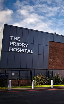
The Priory Hospital, Priory Road, Birmingham, B5 7UG
Chamberlain Clinic, 81 Harborne Road, Edgbaston, Birmingham, B15 3HG
Spire Little Aston Hospital, Little Aston Hall Drive, Sutton Coldfield, B74 3UP
6 Church Street, Oakham, Leicester, LE19 1SJ

Birmingham
Priory Hospital

Edgbaston
Chamberlain Clinic

Sutton Coldfield
Spire Little Aston Hospital

Leicester
Coe & Coe

